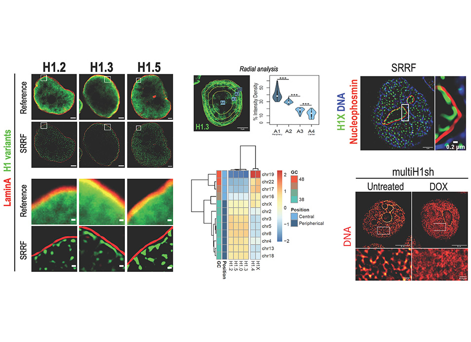
New publication in eLife of the Jordan Lab in collaboration with the IBMB Imaging Platform showing that…
The first article of Cryo-Electron Microscopy Platform has been published on Science
Researchers at the Center for Genomic Regulation (CRG), the National Cancer Research Center (CNIO) and the IBMB-CSIC have captured the world’s first high-resolution images of the earliest moments of microtubule formation inside human cells. The findings, published today in the journal Science, lay the foundations for potential breakthroughs in treating many different types of diseases ranging from cancer to neurodevelopmental disorders.
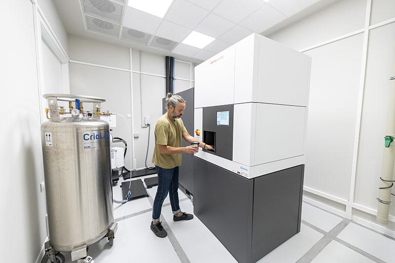
To observe γ-TuRC while it was actively forming microtubules, researchers prepared samples at the CRG and the Cryo-Electron Microscopy Platform of the IBMB-CSIC, located at the Joint Electron Microscopy Center (JEMCA) within the ALBA Synchrotron, where they were flash-frozen in a thin layer of ice – preserving the natural shape of the molecules involved and helping discern fine details of structures at near atomic level. Pablo Guerra, head of the cryoEM Platform at the IBMB-CSIC check all the samples and acquired preliminary data used to evaluate the conditions forming γ-TuRC using a cryo-electron microscope.
Reference:
BRITO C, SERNA M, GUERRA P, LLORCA O,SURREY T.
Transition of human γ-tubulin ring complex into a closed conformation during microtubule nucleation.
Science, eadk6160. DOI: https://doi.org/10.1126/

Related Posts