New publication in eLife of the Jordan Lab in collaboration with the IBMB Imaging Platform showing that…
Expansion microscopy of the chick embryo neural tube to overcome molecular crowding at the centrosomes-cilia
Abstract
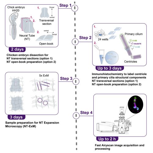
We describe an optimized protocol for application of expansion microscopy (ExM) on chick neural tube (NT) which enables different oriented nanoscale resolution imaging of the centrosomes/cilia. We explain embryo NT transversal sections and open-book preparations, immunohistochemistry for labeling, and sample preparation for 5-fold tissue expansion. Further, we detail sample orientation and Fast Airyscan confocal acquisition and show that NT-ExM retains fluorescence signals and overcomes biomolecules crowding in structural features that to date were only imaged with electron microscopy on tissues.
Reference:
Wilmerding, A., Espana-Bonilla, P., Giakoumakis, N. N., & Saade, M. (2023). Expansion microscopy of the chick embryo neural tube to overcome molecular crowding at the centrosomes-cilia. STAR Protocols, 4(1), 101997. doi: 10.1016/j.xpro.2022.101997


